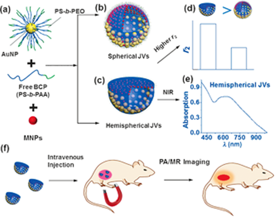
Magneto-plasmonic Janus vesicles (JVs) integrated with gold nanoparticles (AuNPs) and magnetic NPs (MNPs) were prepared asymmetrically in the membrane for in vivo cancer imaging. The hybrid JVs were produced by coassembling a mixture of hydrophobic MNPs, free amphiphilic block copolymers (BCPs), and AuNPs tethered with amphiphilic BCPs. Depending on the size and content of NPs, the JVs acquired spherical or hemispherical shapes. Among them, hemispherical JVs containing 50 nm AuNPs and 15 nm MNPs showed a strong absorption in the near-infrared (NIR) window and enhanced the transverse relaxation (T2) contrast effect, as a result of the ordering and dense packing of AuNPs and MNPs in the membrane. The magneto-plasmonic JVs were used as drug delivery vehicles, from which the release of a payload can be triggered by NIR light and the release rate can be modulated by a magnetic field. Moreover, the JVs were applied as imaging agents for in vivo bimodal photoacoustic (PA) and magnetic resonance (MR) imaging of tumors by intravenous injection. With an external magnetic field, the accumulation of the JVs in tumors was significantly increased, leading to a signal enhancement of approximately 2–3 times in the PA and MR imaging, compared with control groups without a magnetic field.